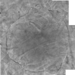
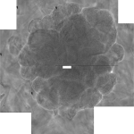
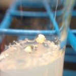
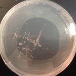
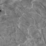
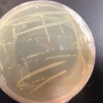
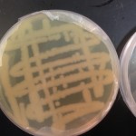
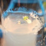
WT E. coli colony (on D2O LB agar) morphology

Yesterday I posted some pictures of E. coli colony morphologies. This was one of the colonies, but it wasn’t as developed, so today I’m adding the extra day’s growth image.

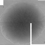
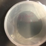
WT E. coli grown on DI LB agar

Looks great! It’s interesting to note that the colonies grown on D2O agar grow out. Instead of getting thick like it normally does, it grows in an outward direction. I guess I would attribute that to the stress induced by being in D2O.

Comparing the results from today to WT E. coli grown on  DI media and D2O adapted E. coli grown on D2O media, it seems there is an interesting mix of morphological behavior. The adapted E. coli is very “brainy” and obviously the normal WT is “smooth,” but today’s specimen is in between smooth and brainy. Unfortunately I can’t make out the topographical features because the E. coli (as I mentioned above) is very flat. But the contour is very feature rich.

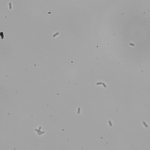
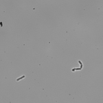
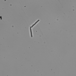
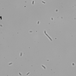
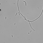
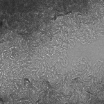
E. coli cells in D2O

I’m not going to make many comments about these cells. It seems that in D2O, E. coli is more likely to say fused but it’s not as obvious as it is with yeast. I make no other observations.

Last Arabidopsis Update

The media is dwindling and the plants will die shortly. I’ll be restarting this experiment in larger environments with more media. Hopefully the plants can do better.

Yesterday Koch noticed that the roots in 10% D2O were much longer than those of the plants in DDW. Something interesting.

E. Coli Colony Morphology in D2O

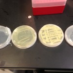
Here are the results of yesterday’s setup. Here I’m comparing 4 samples:

  1. Wild type (WT) E. coli grown on DI LB agar
  2. WT E. coli grown on D2O LB agar
  3. D2O adapted E. coli grown on DI LB agar
  4. D2O adapted E. coli grown on D2O LB agar

All 4 samples were incubated for the same amount of time, and taken from starter cultures of similar absorbance. The absorbances of the starter cultures are as follows:

  1. Wild type (WT) E. coli grown in DI LB – 0.641
  2. WT E. coli grown in D2O LB – 0.325
  3. D2O adapted E. coli grown in DI LB – 0.489
  4. D2O adapted E. coli grown in D2O LB – 0.112

The D2O adapted E. coli took over the DI LB plate! I’ve re-inoculated those cells from that plate to get a picture that would show a typical colony and compare that morphology to the rest. I’ve also allowed the two D2O media samples to incubate for another 24 hours.

It should also be said that the D2O adapted colonies grown on D2O media look distressed compared to the WT colonies on DI media. But compare the two D2O adapted colonies and it’s tough to discern. Whatever mechanism gives the colonies a distressed appearance on D2O media, seems to be completely uninhibited on DI media. It’s tough to tell if the cells are distressed or just out of control.

Still these results seem pretty comparable to the last time I did these experiments (except using YPD). I’ll update again tomorrow, and insert these results into my dissertation.

Preliminary Results of YPD deterioration

Absorbance of DI YPD (pink), D2O YPD (green), and blank (red)

These are the results of the experiment I stated a couple weeks ago. I have been tracking the deterioration (previously called aggregation, but I’m not entirely sure aggregation is the correct terminology) of YPD in both solvents. Today they looked pretty well degraded so I thought I’d share the results. Between the two, the DI YPD is more absorbent than the D2O YPD at nearly every wavelength measure (major uncertainty below 350nm).
I’m associating degradation with absorbance since the blank (which is also DI YPD) has an absorbance of zero at all the same frequencies.

D2O YPD also records 0 for absorbance at 600nm, which is the wavelength used for cell count studies, so there would be no interference from the solution. Whether or not the media is still usable by cells is undetermined.

I’m beginning a second experiment that would track the absorbance every few days via the same mechanism. If you recall, I began this experiment taking pictures and eventually moved toward using the nanodrop. This probe seems to do a good job so its continued use is reasonable.

Man I’ve been writing my dissertation for too long…

D2O Adapted E. coli Experimental Replication

Yesterday while writing I realized that the images of the D2O adapted E. coli that I’ve taken were grown on D2O YPD. In an effort to figure out if the morphologies are due to the YPD or the D2O, I’ve decided to redo the experiments on LB agar.

Today I made some D2O and DI LB broth:

  • 1.84g LB in 92ml of D2O
  • 1g LB in 50ml of DI water
  • Filtered broth for sterilization

Then from there I made some LB agar

  • 40ml of liquid D2O LB with 0.8g of agar (2% agar)

I already have solid LB plates with normal water (commercial).

I then incubated E. coli at 37C in liquid media so that I can streak the plates and analyze them. I used 2 different E. coli:

  1. Normal competent cells
  2. Cells from Day 33.
  3. I also had made a separate culture from an unlabeled glycerol stock that I’m pretty sure I made when I finished the experiment. I’ll check it out tomorrow after the sample has developed.

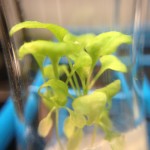
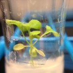
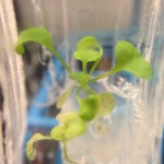
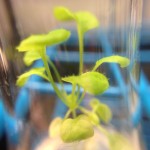
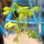
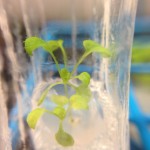
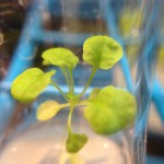
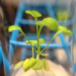
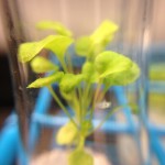
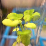
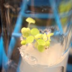
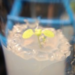
Arabidopsis Update (From March 6, 2013)

I took these images several days ago and again forgot to post them. Stupid dissertation…

Anywho, here they are. I’ll try and remember to update tomorrow when I take the images. Tomorrow will be the last day I update this experiment. I’ll be going into full dissertation mode and will be starting a new experiment when I return. I bought some larger test tubes (1in wide), which should give the plants all the room they need and should be large enough to provide more media to keep the plants alive for longer.

Chapter 1: Open Notebook Science

View in Google Drive.

The link above should give you access to the chapter in all it’s glory. Currently it is pretty much done barring revisions, the addition of figures, and moving the references from side comments to an end of chapter reference section. I’m providing an embed below in case you don’t care about all the cool references enclosed and just want to read. If you are reading via mobile, click the link.

The entire story of my scientific career

This article is actually the introduction to my dissertation and I thought I’d share it with the world officially rather than let it die in an electronic archive somewhere. I’ve shared this story in some form or another several times already, but I’ve never provided the entire account like this. And so, it is with great pleasure that I share with you, the story of how I became the scientist that I am today…

I joined the KochLab in the Spring of 2007. It was a brand new lab that, at the time, was comprised of Dr. Koch, myself, and my best friend Larry Herskowitz (who is now Dr. Herskowitz). In our first lab meeting, Dr. Koch discussed his scientific endeavors up to that point (some of which are continued in this dissertation) and introduced the concept of open science.

Open science was, and still is, an emerging paradigm, and is not to be confused with a particular field of science. The core concept of open science is providing access information and it is through the opening of scientific research that many new endeavors have become possible. Many of these endeavors have changed the way scientists approach research and acquire data. Citizen science, for instance, has brought a mass scale of human analysis to previously unsolvable problems. Even sharing data has led to new forms of collaboration. Data repositories have allowed scientists to share data with the world in hopes of finding new uses for the shared data. Tools like DataOne have emerged to provide some organization to the new data. Meanwhile, open notebook science has emerged to open the entire scientific process and practitioners make every stage of research accessible including protocols, raw data, data analysis, and much more open to scrutiny.
Continue reading The entire story of my scientific career

Do you?

%d bloggers like this: